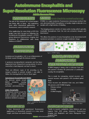
Autoimmune Encephalitis and Super Resolution Fluorescence ‐ Microscopy
Adrián barreros pérez
The aim of this research as a school project was being able to know and experience more about biomedical applications of fluorescence used in scientific research. After applying for some help to ICFO this project was then focused on following the Autoimmune Encephalitis research of ICFO group Advanced Fluorescence Imaging and Biophysics in collaboration with Josep Dalmau at IDIBAPS.
Autoimmune Encephalitis (AE) is an inflammation of the brain caused through the immune system. It produces neuropsychiatric symptoms and has been diagnosed most frequently in young women. It can be
treated but a bad diagnosis can lead to death. There are different types of AE attending to the synapse receptor or protein attacked. I was able to follow the investigations of two of these types:
• Autoimmune Encephalitis AntiNMDA: the immune system attacks NMDA receptors, causing psychiatric symptoms, confusion and memory loss.
• Limbic Encephalitis: the immune system attacks the synaptic protein LGI1 which makes a synaptic port between ADAM22 ADM 23 receptors.
STORM is a superresolution fluorescence microscopy method that uses photoswitchable fluorophores to localize single molecules in a sample with a high density of labeling. The imaging process consists of many
cycles during which optically resolvable fluorophores from the rest are activated, imaged and deactivated.
This allows the position of every fluorophore to be determined with nanometer precision to construct a superresolution image.
STORM imaging is being used to discover how and why patients develop the production of antibodies causing
the encephalitis. This is made by comparing normal neurons and treated neurons with patient’s cerebral
spinal fluid. These neurons are obtained from rats and follow this process: primary culture, staining, STORM
imaging.


